Epidermophyton floccosum (mould/dermatophyte)
Note: While I've had photos of Epidermophyton floccosum for some time now, I've never been satisfied with the quality of the microphotographs I've taken. I remain unsatisfied here. The photos for this and every other post contained in this blog were taken by myself on my own time, before or after regular work hours. Unfortunately I find myself so busy at times that my own projects take a distant back seat. Primary cultures sometimes may become contaminated or overgrown. Isolates may revert to a sterile form on repeated subculture, as in this case, before sufficient study. While I've obtained several isolates over the years, E.floccosum always seems to defeat my best efforts to obtain those "text book" quality photos. Time is running out...
Ecology:
Epidermophyton floccosum is a
cosmopolitan (worldwide distribution)
anthropophilic (man is the primary host
& reservoir) dermatophyte [i]
. A once though related species, Epidermophyton stockdaleae has been
determined to be a synonym for Trhycophyton
ajelloi which exhibits no known pathogenicity to humans or animals
Pathogenicity:
E.floccosum
causes tinea pedis (athlete’s foot), tinea cruris (groin infections or
“jock-itch), and tinea corpis (body infections), and to a lesser extent
onychomycosis (nail infections). Skin infections are also known as “ring-worm”
though there is no ‘worm’ involved.
Infection with E.floccosum may
be transmitted in gym facilities where unprotected feet may share a common
floor. E.floccosum rarely infects the
scalp and does not infect hair or hair follicles.
Macroscopic
Morphology:
E.floccosum exhibits moderate growth, becoming mature in about 10 –
14 days. Surface colonies (media
influenced) have been described as mustard yellow or yellowish brown to
olive-grey (khaki) in colour. Colonies
can be powdery, velvety or felty in texture and acquire a folded appearance as
growth progresses. After prolonged
incubation, sterile floccose (hairy)
white mycelia may cover the colony. The
reverse has been described as ochre, mustard-yellow to yellow-brown and even orange
in colour.

E.floccosum -colony heaped up at center on SAB after 2 weeks at 30ᵒC (Nikon)

E.floccosum -colony on SAB (Saboraud Dextrose Agar) after 2 weeks at 30ᵒC (Nikon)

E.floccosum -colony on SAB after 3 weeks at 30ᵒC (Nikon)

E.floccosum -another colony on SAB after 3 weeks at 30ᵒC (Nikon)

E.floccosum -yet another colony on SAB after 5 weeks at 30ᵒC.
Note: white floccose patches beginning to develop. (Nikon)

E.floccosum -colony on SAB, 30ᵒC after repeated subcultures has developed white floccose patches which are areas of sterile hyphae. (Nikon)

Microscopic
Morphology:
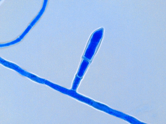
E.floccosum has septate hyphae however microconidia are not
produced which differentiates it from the other genera of dermatophytes. Macroconidia develop as lateral or terminal
outgrowths from mature hyphae and initially lacks a basal septum. Rather thin
walled macroconidia (10 -40 µm X 6 – 12 µm) contain 2 to 5 cells can occur singly
or in characteristic clusters. As the
culture ages, macroconidia may transform into chlamydoconidia (chlamydospores) so
they are best observed earlier in growth. The macroconidia are smooth walled,
and clavate (club shaped) with a blunt tip.
This also differentiates it from Microsporum
and Trichophyton. (Again, see endnote 1).
Note:
Stock cultures are best maintained on SAB
media with 3 – 5% sodium chloride. This
may reduce or prevent the isolate from becoming sterile.

E.floccosum - a first look at low power.
(100X, LPCB, DMD-108)

E.floccosum - Slightly higher magnification reveals the macroconidia more clearly.
(250X, LPCB, DMD-108)

E.floccosum - as above, numerous club shaped macroconidia are clearly seen.
(250X, LPCB, DMD-108)

E.floccosum - club shaped macroconidia with internal septations.
(1000X, LPCB, DMD-108)

E.floccosum - club shaped macroconidia with internal septations.
(1000X, LPCB, DMD-108)
E.floccosum - club shaped macroconidia with internal and basal septations.
(1000+10X, LPCB, DMD-108)

E.floccosum - again, club shaped macroconidia with internal septations. My isolates tended to produce single macroconidia over the grouped macroconidia where several macroconidia crowd each other growing out from the same area of the hypha.
(1000+10X, LPCB, DMD-108)

E.floccosum - a single macroconidium. Note that in this and other microphotographs in of E.floccosum, there are no microconidia.The lack of microconidia is one feature which distinguishes E.floccosum from other dermatophytes.
(1000+10X, LPCB, DMD-108)

E.floccosum - several macroconidia.
(1000X, LPCB, DMD-108)

E.floccosum - a single mature macroconidium.
(1000+10X, LPCB, DMD-108)

E.floccosum - a single mature macroconidium with a curious little kink in it's side.
(1000+10X, LPCB, DMD-108)

E.floccosum - macroconidium measures 35.18 µm in length.
This was obviously an adhesive tape preparation which may trap air bubbles or even reveal uneven adhesive application which may detract from the photograph.
(1000+10X, LPCB, DMD-108)

E.floccosum - club shaped macroconidia. I have this photo recorded as taken at 400X which is confirmed by the micron bar in the upper right. However, the macroconidia seem extremely large for this magnification if compared to previous photos at 1000X. The same goes for the photo which follows. Curious...
(400X, LPCB, DMD-108)

E.floccosum - numerous club shaped macroconidia as above.
(400X, LPCB, DMD-108)
E.floccosum - this photo was taken from a culture that was just over three weeks old. Numerous roundish chlamydospores have developed. Again, compare the micron bar in the upper right to the previous photo which shows identical magnification yet the macroconidia vary greatly is size.
(400X, LPCB, DMD-108)

E.floccosum - macroconidia and chlamydospores present in this adhesive tape preparation
(1000X, LPCB, DMD-108)

E.floccosum - macroconidia on prolonged culture. Some sources say that arthroconidia may also develop, however, I have never observed them in my older E.floccosum cultures.
(500X, LPCB, Nikon)

E.floccosum - finger-like group of macroconidia.
(1000X, LPCB, Nikon)
[i] Dermatophyte – fungi which thrive on
keratin for growth therefore they primarily infect skin, hair and nails
depending on the genera and species. Epidermophyton, Microsporum and Trichophyton
are dermatophytes. Epidermophyton had macroconidia that are clavate (club shaped)
while Microsporum produces fusiform
(spindle shaped) macroconidia and Trichophyton
possesses cylindrical or ‘cigar-shaped’ macroconidia. E.floccosum
does not produce microconida which also serves to differentiate it from the
other dermatophytes.
* * *

.jpg)


