Monday 31 January 2011

Phialophora varrucosa

Phialophora varrucosa
Fungus (Black Mould)

One of the agents responsible for chromoblastomycosis, a slow moving fungal infection of the skin and subcutaneous tissue, usually initiated a splinter or thorn. A cosmopolitan saprobe commonly isolated from soil and decomposing wood. This infection is more common in tropical or sub-tropical climates where it can difficult to treat but is rarely fatal.
Other agents causing chromoblastomycosis are;
-Fonseca pedrosoi
-Cladosporum carrionii
-Fonseca compacta

Growth;
Exhibits slow to moderate growth, usually maturing in about 7 – 12 Days.

Macroscopic Appearance;
Colonies are woolly to velvety, dark grey, brown or olivacious black on the surface and reverse.

SAB agar at 10 Days incubation
(Click on any photo that follows to enlarge for better viewing)

Microscopic Appearance;
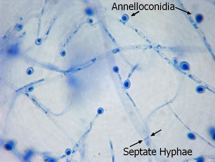
Dematiacious (melanin pigment) - Hyaline to brown, septate hyphae.
Phialides are pale brown to brown, bottle or vase or shaped with a darker collarette at the apical end. Phaialides are located laterally or terminally on the hyphae.
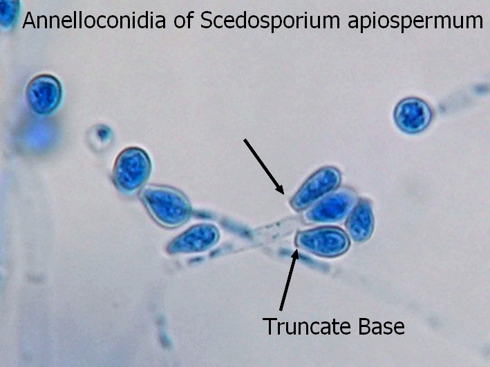
Conidia are unicellular, smooth and thin walled, hyaline to brown and round or ovoid (1-3 X 2-4 µm) which accumulate at the apex of the collarette giving the appearance of a vase of flowers.


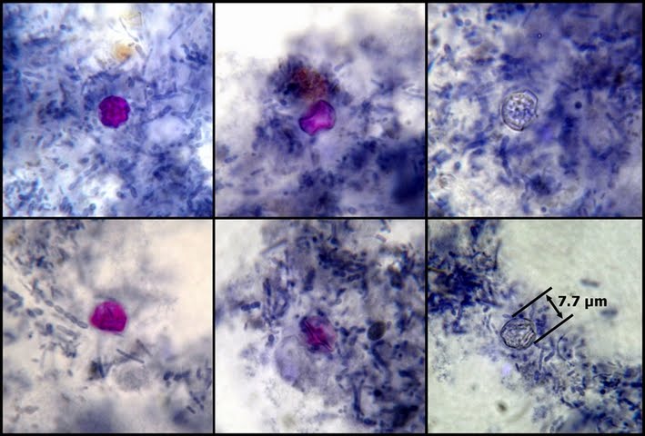
In chromoblastomycosis (histologic specimen), the organisms appear as dark round cells (copper pennies) 5 – 12 µm dia.

Phialophora verrucosa is distinguished from other fungi by its unique phialides with their collarettes. The shape of the collarettes themselves distinguishes Phialophora verrucosa from Phialophora richardssiae, the former being cup shaped, the latter having a flatter, saucer shape.


Vase shaped phialides with round to oval conida at apex. Dark brown (containing melanin) cup shaped collarette at apex. Hyaline to brown (dematiacious ie. containing melanin), septate hyphae,

Ditto

(Click on any photo to enlarge for better viewing)

Vase or bottle shaped phialide showing dark collarette with oval conidia

Voriconazole, Itraconazole with 5-flucytosine have has shown to be effective in treating this difficult infection. Cryosurgery has also been employed.

To compare to Phialophora richardsiae (now known as Pleurostomo richardsiae) click Here.
* * *

Monday 17 January 2011

Pseudallescheria boydii/Scedosporium apiospermum

Pseudallescheria boydii/Scedosporium apiospermum
Fungus
Pseudallescheria boydii (sexual state) -Teliomorph
Scedosporium apiospermum (asexual state)

The names above refer to the same fungus exhibiting different reproductive states. The sexual state name (Pseudallescheria boydii) takes priority over the asexual state name (Scedosporium apiospermum). However, the sexual state often is not present in clinical specimens and unless it is observed, the fungus must be referred to by the asexual name of Scedosporium apiospermum.

Ecology;
The fungus is cosmopolitan and can be isolated from rural soils, polluted water, composts and manure.

Macroscopic (Colony);
  • Moderately rapid growing colony at 25C which matures in about a week.
  • Texture is woolly to cottony.
  • Colony is white becoming greyish or smokey-brown on the surface; reverse is pale with brownish-black zones.
  • Sexual state P. boydii is inhibited by cycloheximide (fungal selection agars) while the asexual state S.apiospermum is not inhibited.

Microscopic;
  • Hyphae are hyaline, septate (2 - 4 µm dia.)
  • Conidiophores bearing annellides are of varying length and exhibit little differentiation from the vegetative hyphae.
  • Conidia (annelloconidia) are unicellular, pale brown, ovoid with truncate bases formed singly or in small clusters at the ends of the conidiophores or from short annellidic necks arising directly from the hyphae.
  • Fascicles of conidiophores bound together in synemata are sometimes present and is a variation of the asexual state referred to as the Graphium synanamorph (one anamorph somewhat resembles a wheat sheaf, the other anamorph a bulb on a stalk[ not flattend as in LPCB prep.]).
  • Large (50 to 250 µm) brown cleistiothecia (of the sexual P.boydii) may develop after 2 to 3 weeks of incubation. More likely to be found at center of colony.
  • Ascospores are yellow-brown and ellipsoidal in shape.
Scedosporium apiospermum hyphae & (annello)conidia
(LPCB sticky tape preparation at 250X)
-click to enlarge-

Scedosporium apiospermum septate hyphae, conidiophores & conidia
(LPCB sticky tape preparation at 400X)
-click to enlarge-

Annelloconidia showing truncate base/scar -wider at apex
LPCB 500X
-click to enlarge-

Scedosporium apiospermum asexual Graphium synanamporph
(Fascicles of conidiophores bound together in synemata -wheat sheaf appearance)
-click to enlarge-

Graphium synanamorph
(Slide culture on Corn Meal Agar-edge of agar at photo bottom 250X)
-click to enlarge-

As Above Description
-click to enlarge-

Immature Cleistothecium at 10 days Corn Meal slide culture LPCB
(Hyphae & Conidia can be seen in surrounding background)
-click to enlarge-

Immature Cleistiothecium of Pseudallescheria boydii
(Corn meal agar slide culture-unstained X400 ~10 days)
-click to enlarge for better viewing-

**New: Feb 17th, 2011

Split Mature P.boydi clestiothecium
(taken with the DMD-108 microscope)
(click on image to enlarge)

Mature P.boydi clestiothecium split and releasing ascospores
(ascospore inset)
(Click on photo to enlarge for better viewing)

P.boydi clestiothecium split open revealing lemon shaped ascospores within.
(click on photo to enlarge for better viewing)

***Note: The sexual stage of P.boydi was induced using corn meal agar both as a slide culture and extended incubation of the plate media. The last two photos (Feb 17th) from corn meal agar media after about 8 weeks of incubation. A small segment of agar was cut out of the plate and placed on a glass microscope slide. A second slide was placed on top of the agar and the agar was crushed between the two (The clestiothecia of P.boydi were much more difficult to split open than those of Aspergillus nidulans.) The the two slides were then separated, leaving material on both. A drop of KOH was added to help clear and define the organism.

Pathogenicity;
An occasional agent of infections including mycetoma, cutaneous or subcutaneous invasion, most often of the feet and hands. Has been implicated in otitis, sinusitis, keratitis, endophthamitis, pneumonia, endocarditic, meningitis, osteomyelitis and cerebral abscesses. Disseminated disease more likely to occur in debilitated patients or those immunocompromised. Is also now recognized as an agent of phaeaohyphomycosis

Susceptibility;
Reference

* * *

Sunday 9 January 2011

Cyclospora cayetanensis

Cyclospora cayetanensis
(Cyanobacterium-Like Body/Coccidian-Like Body/CLB)
Coccidian Protozoan Parasite

Though the genera Cyclospora has been known about for some time as pathogen of vertebrates, this nasty little species began to be increasingly reported in humans in the late 1970’s. First thought to be associated with blue-green algae, it was referred to as a Cyanobacterium-Like body. Others thought it was a larger variant of Cryptpsporidum. The organism was finally identified as belonging to the genus Cyclospora based on sporulation studies which revealed that each oocyst contains two sporocysts which in turn contain two sporozoites.

Cyclospora can be found worldwide however it may exhibit greater prevalence in rainy areas where outbreaks coincide with the rainy season. Infection is primarily acquired by ingestion of water bearing the oocyst or produce which has been irrigated with contaminated water. There is no proof of person to person infection as the oocyst needs about 10 days outside of the body to mature and become infective.

On ingestion, the oocyst takes up residence within cells (enterocytes) of the small intestine. Cyclospora appears to have both asexual and sexual reproduction with the infective oocyst being the product of sexual reproduction. The invasion of the intestinal enterocytes is thought to stimulate cytokine production which causes inflammation, electrolyte secretion and ultimately osmotic diarrhoea.

Otherwise healthy individuals may suffer a prolonged, yet self-limiting watery diarrhoea. Symptoms may include vomiting, nausea, fatigue, anorexia, fever, cramping and weight loss. Episodes of diarrhoea may alternate with constipation. Symptoms may begin 2 to 10 days after exposure and may last up to 7 weeks.
In immunocompromised patients such as those with AIDs, the illness can persist longer and have devastating consequences.

Diagnosis and Identification;
The organism is shed irregularly so stool specimens should be collected periodically if Cyclspora is suspected. The organism can be identified by several means including;

  • Wet-preps of formalin-ether concentrates - the spherical organisms are about 8 to 12 µm and appear refractile in unstained preparations.
  • Cyclospora exhibits variable acid-fast properties and organisms will stain to various degrees from virtually unstained through increasingly intense shades of pink. Stained oocyst may appear slightly smaller than the unstained 8 to 12 µm due to shrinkage. Cells often display a folded or wrinkled appearance. Cyclospora does not stain well with the Trichrome method.
  • Cyclospora will auto fluoresce under ultra-violet light, appearing greenish under 450 nm wavelength and bluish under 365 nm.
Treatment;
Septra (Trimethoprim-Sulfamethoxazole) is commonly used in treatment.

Note: Cyclospora have been known to survive routine chlorination treatment.

(click on photo to enlarge for better viewing)

Above: Cyclspora cayetanensis as stained by the modified Iron-Hematoxylin staining method.
Organism exhibits variable acid-fastness thereby appearing clear/unstained right through various intensities of pink. Note too the wrinkled or folded appearance the organism can have in the stained preparation. Wrinkling and a slightly smaller diameter may be due to shrinkage during the staining process. (photos X1000 oil immersion)

(click on photo to enlarge for better viewing)

Formalin-ether stool concentrate wet preparation showing unstained Cyclospora cayetanensis.

Return Home
* * *